Oropharyngeal candidiasis (commonly known as oral thrush) is an infectious condition of the mucous membranes initiated by the pathogenic overgrowth of Candida yeast microorganisms. Since the fungus is part of the resident flora (present in up to 90% of healthy individuals), the clinical picture develops exclusively against the background of a localized or systemic immune barrier failure. Typical symptoms range from white curdy patches to fiery hyperemia (redness), accompanied by xerostomia (dry mouth) and dysgeusia (taste distortion). The gold standard for diagnosis is a morning microscopic smear followed by mandatory antifungal susceptibility testing. The therapeutic protocol includes systemic antifungals, full dental sanitation, denture sterilization, and strict dietary restrictions on carbohydrates.
Etiopathogenesis: Mechanisms of Opportunistic Flora Transformation
The infectious process in the oral cavity never occurs without a trigger. The primary catalyst is a critical reduction in tissue barrier function due to inadequate daily oral hygiene. Untreated carious lesions and periodontal inflammation (gingivitis) act as a continuous source of infection.
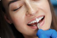
A significant role is played by orthopedic appliances, especially removable dentures. Acrylic and nylon bases have a microporous structure where, if poorly cleaned, a bacterial-fungal biofilm instantly forms. If a patient sleeps with their dentures, a "greenhouse effect" occurs: tissues are deprived of oxygen and natural salivary washing, triggering an explosive growth of anaerobes. Furthermore, epithelial integrity can be compromised by traumatic factors like overhanging filling margins, chipped enamel, thermal burns, or tongue piercings.
Systemically, the disease often manifests as an indicator of severe underlying issues such as HIV, oncological processes, or severe malnutrition. Decompensated diabetes mellitus is a major risk factor: excess glucose in biological fluids (including saliva) creates an ideal nutrient base for the mycelium.
Among iatrogenic (medication-induced) causes, prolonged use of broad-spectrum antibiotics destroys the beneficial microbiome that normally inhibits fungal growth. Immunosuppressants and inhaled corticosteroids for asthma act similarly. Dietarily, a regimen saturated with refined sugars and fast carbohydrates drastically increases the fungus's ability to adhere to epithelial cells.
Clinical Picture and Diagnostic Protocols
Depending on the patient's status and disease duration, dentists distinguish four specific clinical forms:
-
Acute pseudomembranous candidiasis: The most recognizable variant. Loose, whitish, cottage cheese-like patches form on the tissues. Initially, this plaque can be easily wiped off, revealing a bright red, sometimes bleeding surface.
-
Acute atrophic candidiasis: The most frequent consequence of antibiotic therapy. Curdy patches are entirely absent. The mucosa appears thinned, smooth, and fiery red. Patients experience excruciating burning, severe dryness, and metallic or bitter taste alterations.
-
Chronic atrophic candidiasis: Specific to denture wearers (denture stomatitis). Inflammatory reaction, mild edema, and redness are strictly localized to the area where the denture base contacts the gums or palate.
-
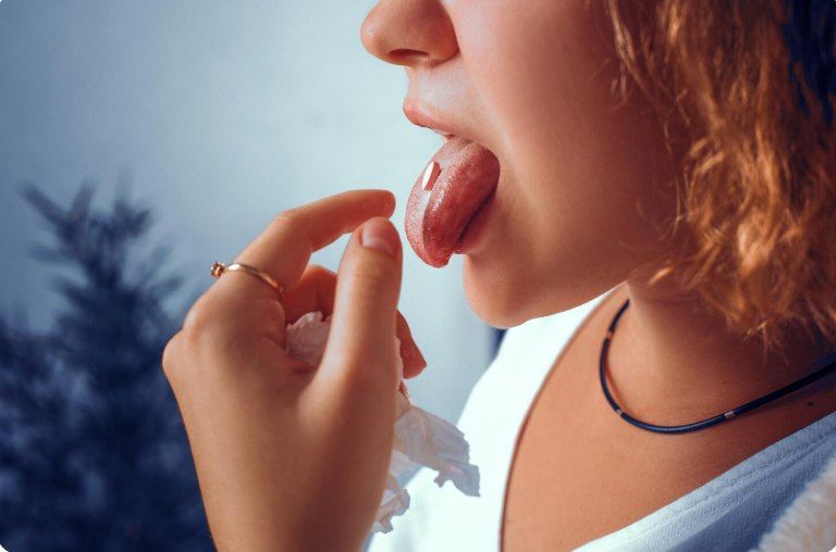
Chronic hyperplastic candidiasis: Typical in older patients and heavy smokers. The infection involves the tongue dorsum, inner cheeks, and palate. Instead of loose plaque, tough, rough, nodular elements form, which may yellow over time. They are firmly fused to the tissues and cannot be scraped off.
Visual examination alone is insufficient for a correct diagnosis. The gold standard is a microbiological swab. It must be taken early in the morning, fasting, and strictly before brushing teeth. A crucial laboratory step is the antifungal susceptibility test to prevent prescribing ineffective drugs to which the strain has developed resistance (e.g., fluconazole resistance is highly prevalent).
Treatment Protocols
Suppression of fungal activity requires a multi-step approach. Systemic antifungal therapy (polyenes, modern imidazole derivatives) is prescribed strictly based on the culture results. Local therapy aims to sanitize tissues and relieve pain, including antiseptic rinses (chlorhexidine, iodine-based solutions), application gels, antifungal lozenges, and probiotics to restore the microbiome.
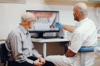
Dental sanitation is mandatory: removal of calculus (tartar), filling of cavities, and extraction of unsalvageable roots. Ill-fitting dentures must be adjusted or replaced. Removable denture wearers must be retaught to disinfect their appliances daily.
Diet is a fundamental part of the therapy. Fast carbohydrates, refined sugar, sweet drinks, and yeast-based pastries are strictly forbidden. Spicy, acidic, or smoked foods that irritate the mucosa are excluded. This dietary regimen must be maintained for 1.5 to 2 months after the clinical symptoms disappear to minimize the risk of relapse.
Expert FAQ
How to distinguish thrush from normal food debris?
Food debris easily washes away with water. Candidiasis colonies form a dense, curdy texture; if scraped with a spatula, an eroded, painful, and red epithelium is exposed underneath.
Can oral thrush be cured with home remedies?
Absolutely not. Baking soda or herbal rinses can temporarily dull symptoms and wash away visible plaque, but they lack fungicidal (fungus-killing) action. The pathogen remains, the disease becomes chronic, and the microorganism gains drug resistance.
Why does the disease often return?
Relapses are usually linked to violating rehabilitation rules: reintroducing sugar too early, failing to replace an old toothbrush, poor denture sterilization, or uncontrolled blood sugar levels in diabetics.
Is it contagious through kissing?
Transmission of the microorganism during close mucosal contact is possible. However, in a healthy partner with a strong local immune system, the infection will not develop.
Can you catch it from shared utensils or toothbrushes?
Using an infected person's toothbrush carries the highest risk: stiff bristles leave micro-injuries on the gums where the pathogen is directly rubbed in. Sharing poorly washed utensils is less likely but possible. The patient must use strictly individual hygiene and eating items.