Teeth Shot
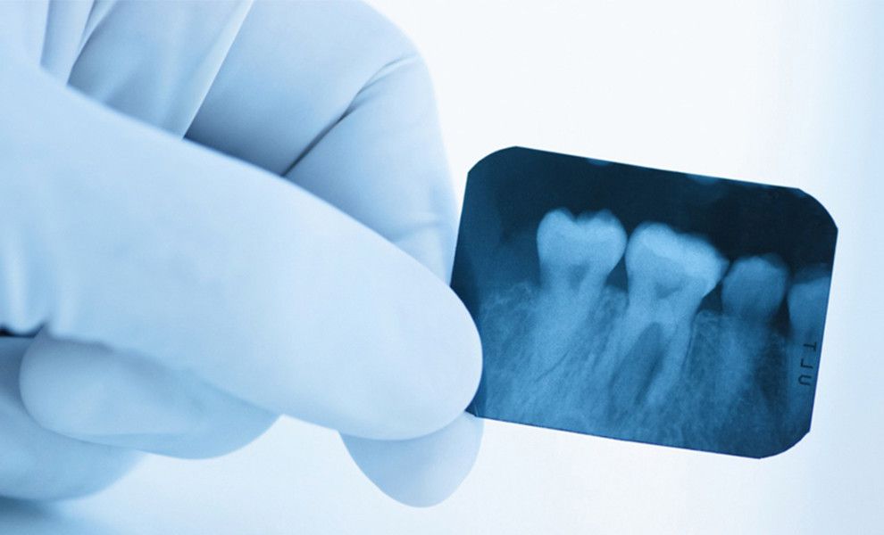
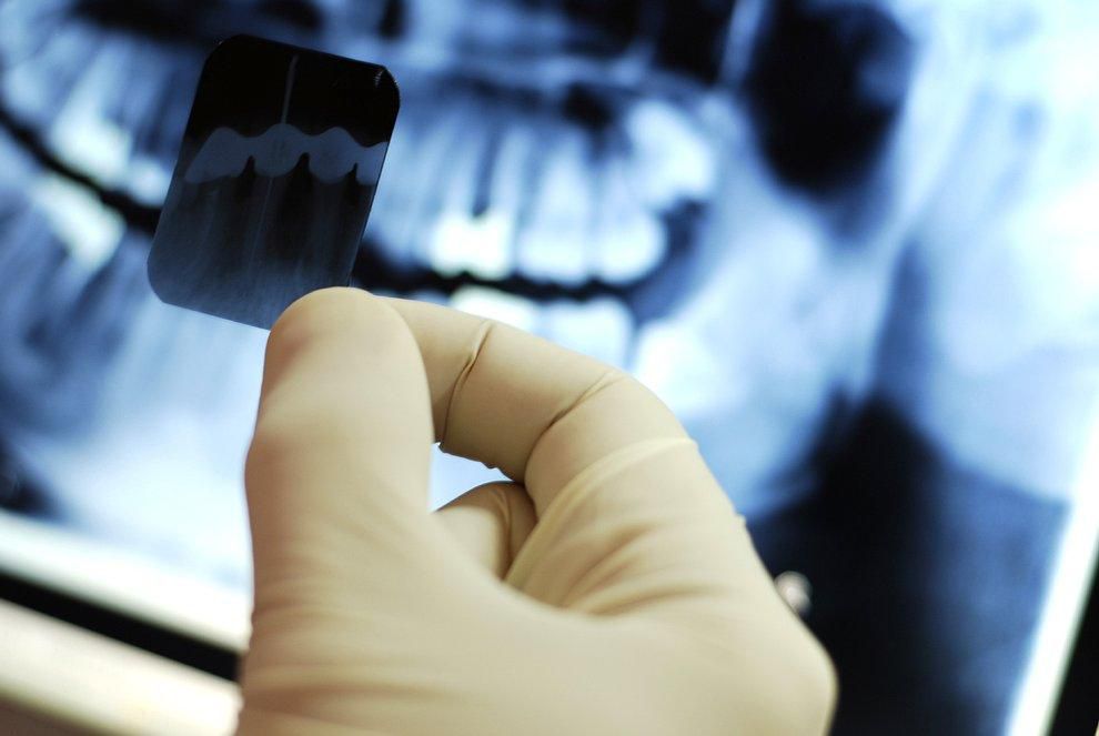
Periapical X-rays
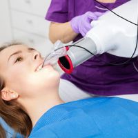
At our clinic, periapical radiographs are obtained using digital radiovisiography.
Contents:
- What is a dental visiograph?
- What is a visiograph used for?
- What are the advantages of a visiograph over conventional X-ray?
- Is dental visiography harmful? Radiation exposure during dental visiography.
What is a dental visiograph?
A dental visiograph (also referred to as a radiovisiograph or RVG) is a modern digital periapical radiographic imaging system.
With conventional radiography, the image is captured on film. In digital visiography, the film is replaced by a digital sensor that transmits data directly to a computer, where dedicated software processes it and displays the image on screen in real time.
This diagnostic modality reduces radiation exposure — for both the patient and the clinician — by a factor of ten.
What is a visiograph used for?
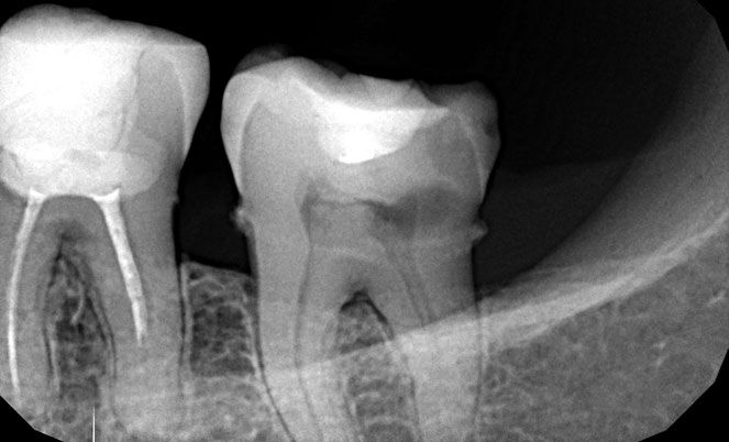
It is used to identify conditions that are not visible on clinical examination:
- Detection of hidden caries and pulpitis.
- Identification of infectious and inflammatory foci (periapical periodontitis, periodontitis).
- Localisation of posts and fractured instruments within root canals.
- Assessment of peri-implant bone status (peri-implantitis).
- Monitoring of each stage of root canal treatment.
- Monitoring of each stage of implant placement.
- And other applications.
What are the advantages of a visiograph over conventional radiography?
- The sensor has significantly greater sensitivity than film, requiring a shorter exposure time to produce an image. This reduces radiation dose compared to standard radiography by up to 90%.
- The image is displayed on a computer monitor with fully adjustable brightness and contrast, and any area of interest can be magnified. This provides far greater diagnostic detail than a conventional film radiograph.
- The minimal radiation dose allows virtually unlimited exposures to be taken, enabling close monitoring of every stage of treatment.
- At our clinic, each surgery is equipped with its own visiograph, meaning that imaging is carried out chairside without interrupting treatment. Patients no longer need to leave the surgery and walk to a separate X-ray room.
- Digital images are stored within the patient's electronic record, making them instantly accessible. They can be reviewed, processed, saved to an external device, or forwarded by email at any time.
Is dental visiography harmful? Radiation exposure during dental visiography.
The permissible effective radiation dose for patients undergoing routine diagnostic radiological examinations is 1,000 µSv per year. Such a dose would only be accumulated through repeated imaging carried out on clinical grounds — for example, multiple CT scans in a single year following severe trauma or accidents. A single periapical exposure taken with a visiograph delivers approximately 2–3 µSv.
This means that, over the course of a year, a patient could safely undergo approximately 330–500 periapical radiographs within the permissible limit.
By way of comparison, the radiation dose received during a flight from London to Moscow is equivalent to roughly half the dose a patient would receive from a CT scan of both jaws. It is also worth bearing in mind that we are all exposed to background radiation on a daily basis — from sunlight, cosmic rays, and naturally occurring sources in the environment such as soil and building materials.
Radiological diagnosis in dentistry therefore poses no meaningful risk to health. On the contrary, it plays an essential role in reaching an accurate diagnosis, planning treatment, and monitoring clinical progress — all of which have a direct and positive impact on treatment outcomes and long-term prognosis.
Contact Information:
| Torrevieja, Pasaje Pais Vasco, edificio 1 local 4 | |
| +(34) 638 893 141 | |
| +(34) 638 893 141 | |
| apdenta@gmail.com | |
| Working hours: Mon - Fri: from 10:00 to 20:00 |