Dental CT Scans Torrevieja Spain
- When does a doctor prescribe a dental computed tomography (CT) scan?
- Dental CT scan before implantation.
- Dental CT scan before root canal treatment.
- Is a dental CT scan harmful? Radiation exposure during a dental CT scan.
- How to prepare for a dental CT scan?
- Specifics of performing a dental CT scan at the "AP-Denta" clinic.
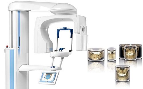
Dental Computed Tomography (3D image, TAC Dental) is the most accurate method of X-ray diagnostics, allowing to obtain a three-dimensional image of the area under study. This type of examination differs from panoramic radiography in that the doctor can examine bone structures, teeth, joints, soft tissues in any projection, from any angle without distortion.
Why is this needed? To make a diagnosis and create a treatment plan, as well as to monitor its quality.
- When does a doctor prescribe a dental computed tomography (CT) scan?
- At the stage of implantation planning.
- In cases of injuries, fractures of the jaws and tooth roots.
- Planning operations related to neoplasms and inflammatory foci of infection.
- Diseases and dysfunctions of the temporomandibular joint.
- Re-treatment of root canals, removal of foreign bodies (broken instruments) from root canals.
- Treatment of inflammatory processes at the apexes of tooth roots ("cysts").
- In severe generalized periodontitis.
- In diseases of the salivary glands.
- During the planning of orthodontic treatment.
- And others.
Dental CT scan before implantation.
In the image, the doctor examines the area where the implants are planned to be placed. There, he studies the condition of the bone tissue, its quality, density, thickness, and width. In a special program, he calculates the diameter and length of the implant, as well as the direction of its placement. He calculates the need for bone grafting. In addition, he pays attention to adjacent anatomical structures: the maxillary sinus, large nerves, and blood vessels.
Understanding the overall picture, the dentist selects the best implantation method and the implant itself. Without a CT scan, the doctor performs implantation at his own risk, endangering the patient!
More details about implantation can be read in the "Implantation" section.
Dental CT scan before root canal treatment.
A CT scan may be necessary before root canal treatment. This examination helps to see the number of root canals, their shape, and location. It also helps to detect hidden inflammations at the root apexes and determine their size.
Read more about root canal treatment in the "Endodontics" section.
Is a dental CT scan harmful? Radiation exposure during a dental CT scan.
The relative permissible effective radiation dose for a patient during preventive radiological examinations is 1000 μSv per year. This dose can be received with regular radiological monitoring for vital indications (several CT scans per year in cases of severe injuries, accidents, etc.).
During a single dental CT procedure on a Planmeca device, the dose ranges from 10 to 70 μSv, an orthopantomogram is approximately 20 μSv, and an intraoral X-ray is approximately 3 μSv.
Consequently, per year, one can have from 14 to 100 dental CT scans, 50 panoramic X-rays, and 330 intraoral X-rays.
For comparison: the radiation exposure during a flight, for example, from Moscow to Spain, is half the radiation dose a patient receives during a CT scan of both jaws. In addition, do not forget about radiation from the sun, cosmic rays, and the environment (soil, building materials, etc.).
Therefore, X-ray diagnostics in dentistry will not harm health; on the contrary, it will contribute to making a diagnosis, allow treatment planning, and monitor the treatment process. This will generally have a positive effect on the treatment outcome and prognosis.
How to prepare for a dental CT scan?
There are no special rules for preparing for a CT scan.
The only requirement is to remove earrings, chains, hairpins, and dentures, i.e., remove all metal objects from the head and neck.
Specifics of performing a dental CT scan at the "AP-Denta" clinic.
Despite the fact that dental CT is the most modern diagnostic method, not all devices are the same, and not all provide an excellent final result.
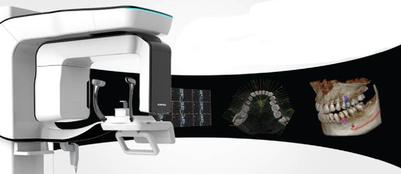
In our clinic, the examination is performed on a Planmeca ProMax® 3D Gen 5 device. This is the newest generation of one of the best tomographs in the world. This model combines the latest developments in X-ray diagnostics and advanced computer programs, allowing for the highest quality and most accurate diagnostics.
This device has a motion artifact correction function, a radiation dose reduction function, a facial scanning and mandibular movement recording function, an endo-mode function (imaging of the smallest details, for example, imaging of a single tooth), and many others.
The unique features of the Planmeca tomograph allow solving absolutely any clinical problems, from identifying hidden carious cavities and endodontic treatment to comprehensive restoration of dentition and creating an ideal smile.
More details about the advantages of this tomograph can be read in the "Technologies - CT-Planmeca Pro Max" section.
In addition, of course, there is convenience for the patient: the examination is carried out in the clinic directly during the consultation, and the results are displayed directly on the doctor's computer screen within a couple of minutes. If necessary, this image can be recorded on a CD or sent to the patient by email.
Contact Information:
| Torrevieja, Pasaje Pais Vasco, edificio 1 local 4 | |
| +(34) 638 893 141 | |
| +(34) 638 893 141 | |
| apdenta@gmail.com | |
| Working hours: Mon - Fri: from 10:00 to 20:00 |